
Longitudinal cortical thickness changes in first episode psychosis: a triple-blind randomised placebo-controlled MRI study
S. Chopra1,2, A. Holmes3, A. Segal4, X. Zhang5, S. Francey1,2, B. O’Donoghue1,2, V. Cropley2,1, B. Nelson2,1, J. Graham2,1, L. Baldwin2,1, H.P. Yuen2,1, K. Allott2,1, M. Alvarez-Jimenez1, S. Harrigan6,2, A. Holmes7, C. Pantelis8, S. Wood2,1, P. McGorry2,1, A. Fornito3
1University of Melbourne, Center for Youth Mental Health, Melbourne, Australia, 2Orygen, Orygen, Melbourne, Australia, 3Monash University, Turner Institute for Brain and Mental Health, Melbourne, Australia, 4Yale University, Neuroscience, New Haven, United States, 5Yale University, Department of Psychology, New Haven, United States, 6University of Melbourne, Melbourne School of Global and Population Health, Melbourne, Australia, 7Rutgers University, Department of Psychiatry, New Jersey, United States, 8University of Melbourne, Department of Psychiatry, Melbourne, Australia
Background: Changes in cortical thickness are a common finding in Magnetic Resonance Imaging (MRI) studies of people with psychosis and have been shown to progress with ongoing illness. A major unresolved question concerns whether these changes are driven by the illness itself or represent iatrogenic effects of antipsychotic medication. This uncertainty has prevented guidance on whether antipsychotic medications potentially protect or harm brain tissue during critical early stages of psychotic disorders.
Aims & Objectives: Our primary aim was to determine whether antipsychotics prevent or cause cortical thinning in first episode psychosis FEP. Our secondary aim was to understand the molecular substrates underlying illness-related cortical thinning.
Method: We report findings from a triple-blind randomised placebo-controlled MRI study where 62 antipsychotic-naïve people with FEP received either an atypical antipsychotic or a placebo pill over a treatment period of 6 months. A healthy control group (n=27) was also recruited. Anatomical T1-weighed scans were collected at baseline, 3-months, and 12-months. Linear mixed effects models were fitted to >150,000 cortical surface locations used to examine illness- and antipsychotic-related changes in cortical thickness. We also examined whether cortical changes were enriched within canonical functional networks or cytoarchitectonic types of laminar differentiation, and spatially correlated with normative receptor/transporter densities measured in vivo via PET imaging.
Results: We found widespread cortical thinning over 12 months (p<.05FDR) in people with FEP receiving placebo compared to healthy controls, with the largest effects [AF1] occurring in frontal, cingulate, and occipital areas. No significant thinning was detected in antipsychotic-treated people with FEP. Highly differentiated koniocortical areas defined using the von Economo and Koskinas atlas were relatively protected from cortical thinning (pspin<.05FDR). Thinning in the placebo group spatially aligned with normative distributions of GABAA/BZ, 5HT1B, 5HT2A, and H3 receptors (.17 < r < .28; pspin<.05FDR). No significant associations between cortical thinning and symptom changes were identified.
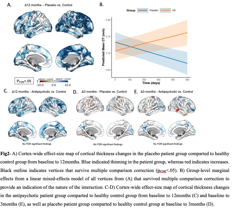
Discussion & Conclusions: Widespread thinning of cortical grey matter occurs over the first year of FEP in the absence of antipsychotic treatment. No such thinning was evidence in patients who received antipsychotics. These findings suggest that cortical thinning in the early stages of psychosis is an illness-related phenomenon that may be mitigated by early antipsychotic exposure.
In sessions